Transmission Electron Microscope
Transmission electron microscope (TEM) has three essential components:
- An electron gun, which produces the electron beam, and the condenser system, which focuses the beam onto the object
- The image-producing system, consisting of the objective lens, movable specimen stage, and intermediate and projector lenses, which focus the electrons passing through the specimen to form a real, highly magnified image
- The image-recording system, which converts the electron image into some form perceptible to the human eye.
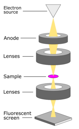
Figure 1. Transmission Electron Microscope
Electrons can interact with the samples in two ways: scattering elastically or inelastically. Elastic scattering produces back-scattered electrons (BSE) whereas inelastic produces secondary electrons (SE). BSEs are incidentally scattered by the specimen atoms, and typically deflect in larger angles with little energy loss. SEs are ejected from the specimen atoms in smaller angles and less energy as kinetic energy is transferred to the specimen atoms. The major difference is that BSEs come from the microscope electron beam, whereas SEs come from the sample itself. BSEs are used for elemental composition. contrast, and SEs for topographic contrast. A vacuum environment is required in a TEM because collisions between high-energy electrons and air molecules significantly absorb electron energy. Other issues include phonon scattering from the atomic lattice vibrations, and the inner shell electrons being ejected (called Auger-electrons). Same theory can be found in the SEM in different wording, and schematic diagram.
TEM can be used to observe particles at a much higher magnification and resolution than can be achieved with a light microscope because wavelength of an electron is much shorter than that of a photon. Resolution in microscopy is limited to about 1/2 of the wavelength of the illumination source (photon/electron beam) used to image the sample. Because our eyes can only detect photons with wavelengths greater than ∼400 nm, the best resolution that can be achieved by light microscopes is about ∼200 nm
Applications of the SEM
- Microstructural analysis of metals and alloys using high-resolution images
- Study of the structure and texture of crystals and metals at molecular level
- Identification of flaws, fractures and damages to micro sized objects